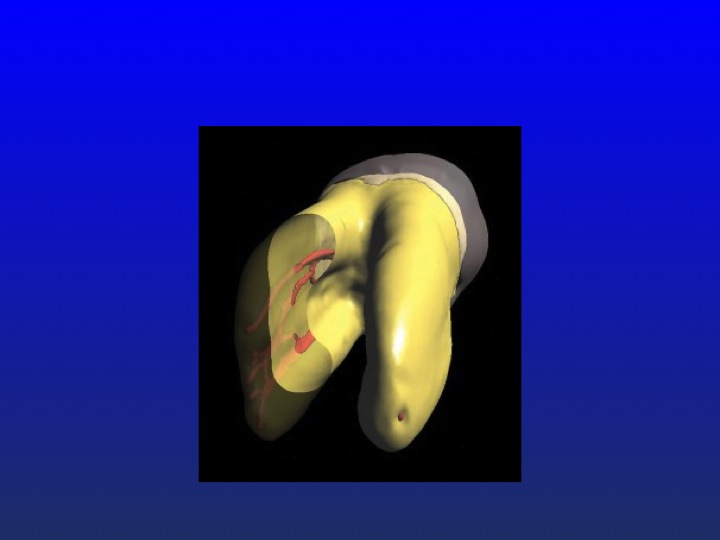
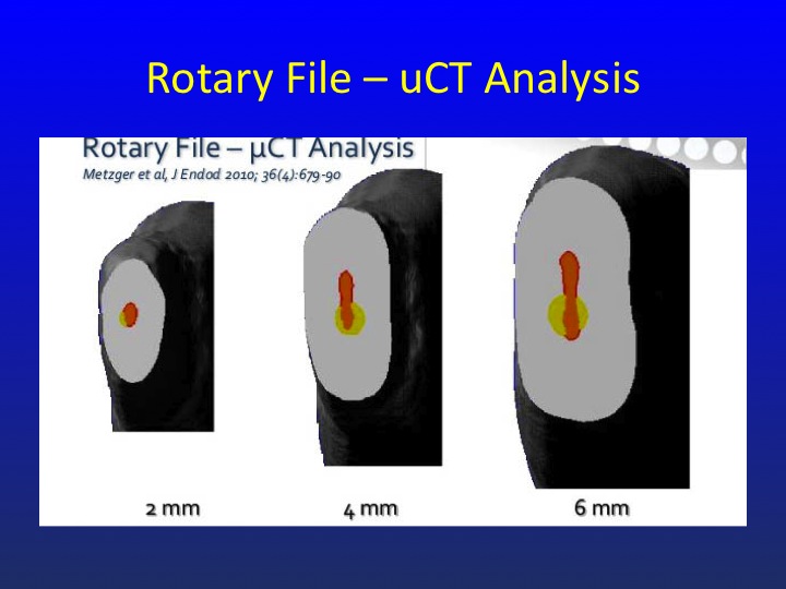
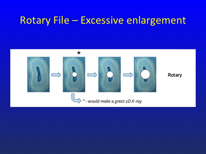
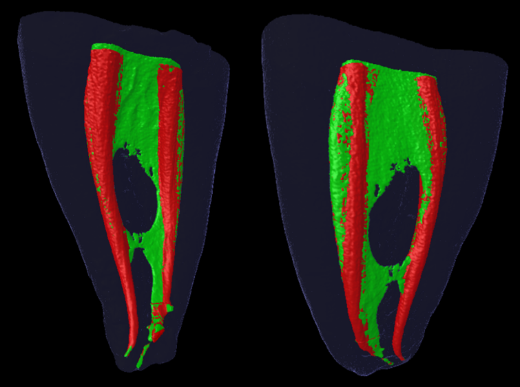
It’s been said many times that one picture is worth a thousand words. When it comes to rotary NiTi preparations of teeth, we now have access to literally thousands of pictures that undermine the concept of conical shaping as a constructive event. Rotary NiTi can make a case for itself in the few situations where original canal anatomy is conical, but those situations are the exception rather than the rule. Below is a small sampling of the imagery that has been produced over the past few years that clearly demonstrates anatomy that is highly oval, sheath-like, extremely thin in the mesio-distal plane and far broader in the bucco-lingual plane, pulpal configurations that are not going to be well debrided because a conical shape has been superimposed. As some of the images below show there is little room for dentin removal in the mesio-distal plane, a fact that is pretty much ignored by greater tapered shaping and further aggravated by the fact that rotary is most safely used when it remains centered, a reality that is a necessity for the instruments but detrimental to the tooth.
After reviewing these images, it becomes obvious that the twin goals of thorough debridement and the conservation of dentin can only be accomplished with thinner instruments that can adapt to the shape of the canal enlarging it more or less uniformly. However, to accomplish this task the dentist must be confident that the instruments will remain intact as they are vigorously applied to the tissue and walls in the bucco-lingual plane. The most obvious way to maintain intact instruments is to avoid their exposure to excessive torsional stress and cyclic fatigue, something that routinely occurs when instruments are used in full rotations whether continuously or in an interrupted fashion. 30º-45º reciprocation virtually eliminates excessive torsional stress and cyclic fatigue and gives the dentist the confidence to apply 02 tapered relieved reamers knowing they will remain intact. As an additional source of confidence, neither straight-line access nor preflaring is a prerequisite for their safe usage.
Please look at the images below. They tell a story that if we listen is directing us away from greater tapers primarily for the safety of the tooth, but also for the instruments we must employ.
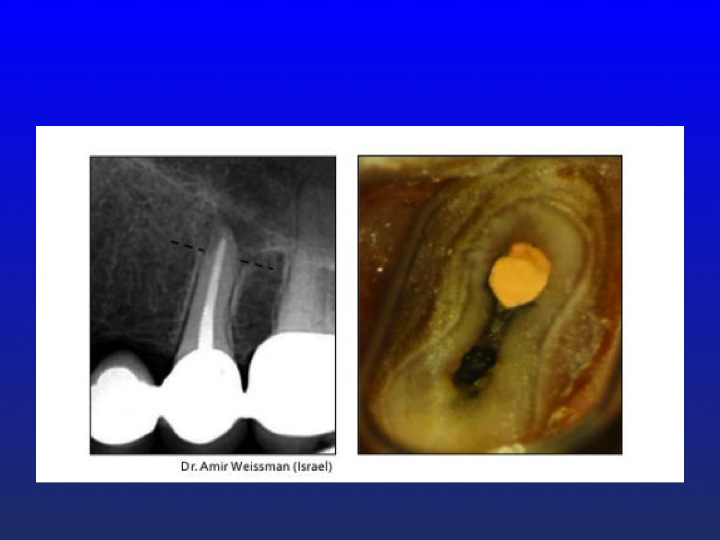
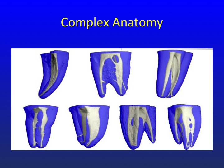
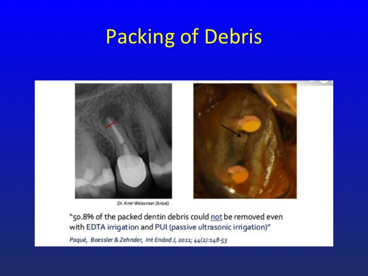
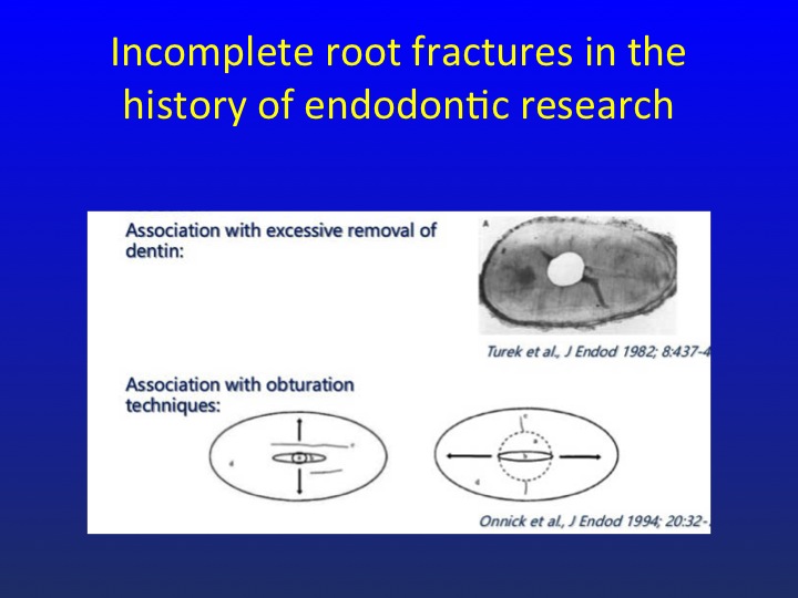
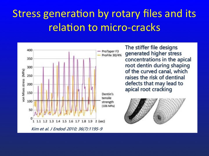
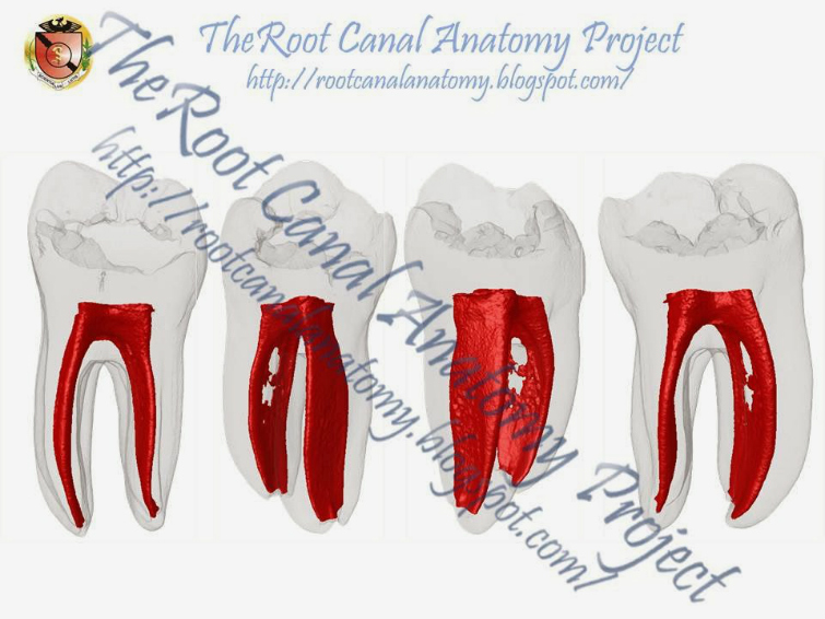
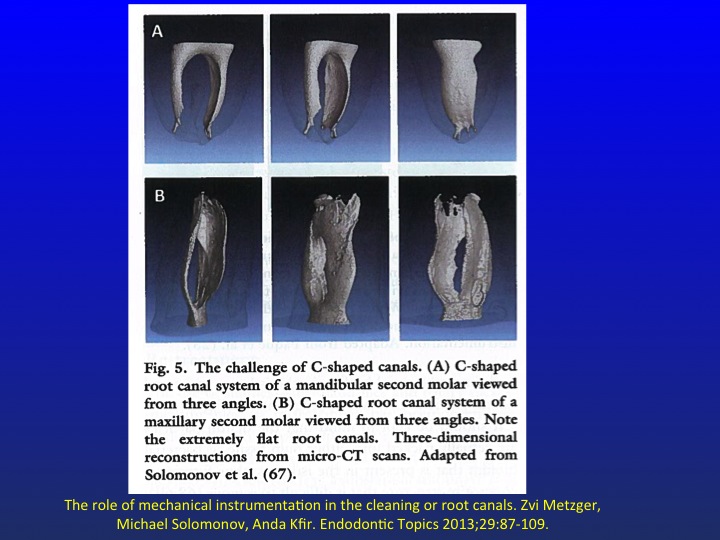
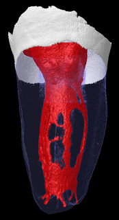
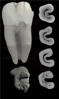
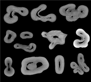
I think there is enough imagery here for us to gain a more accurate perspective of what pulpal anatomy can often look like and drive home the point that for the most part conical shaping is a pleasant configuration that often bears little resemblance to reality. If we don’t ground ourselves in a more accurate view of what we are attempting to cleanse we will continue to remove excess tooth structure in the tooth’s narrow plane, inadquate tissue removal in the broader plane while weakening the tooth in the process and subjecting the instruments to stresses that would be best avoided.
Regards, Barry